
TL;DR
Indian scientists have created the most detailed 3D atlas of the human brainstem to date. This breakthrough enhances understanding of brain anatomy and could impact neurological research and treatment.
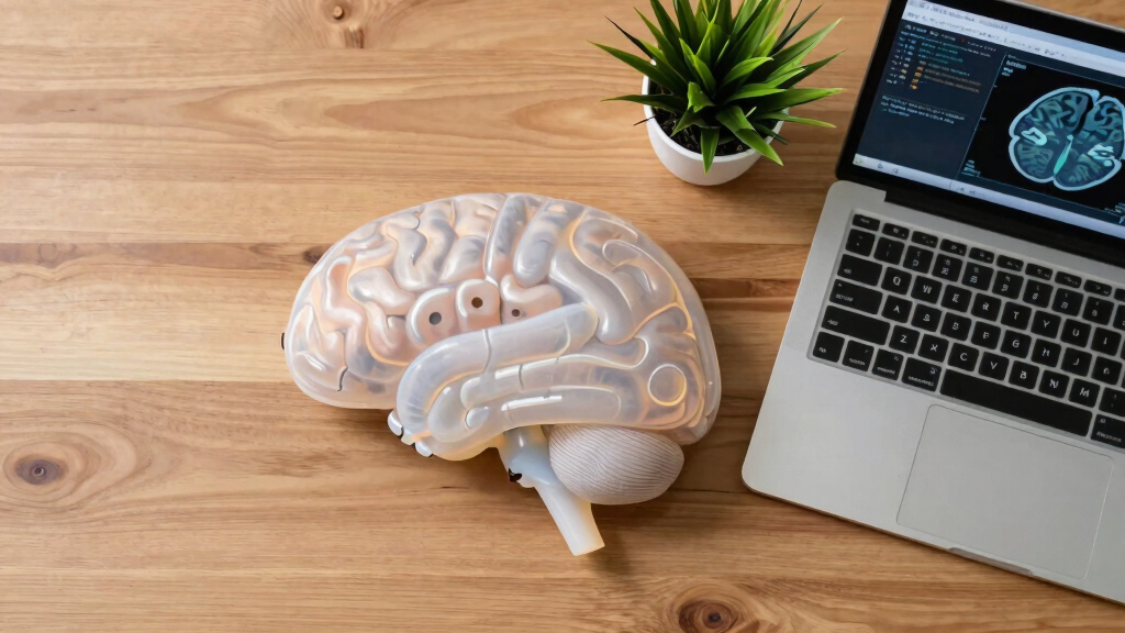
Indian scientists have unveiled the most detailed three-dimensional (3D) atlas of the human brainstem, a development that could contribute to advances in neuroanatomy and medical imaging. The new atlas provides high-resolution detail, enabling researchers and clinicians to better understand the structure of this brain region.
The research team, led by scientists at the Indian Institute of Science, used advanced imaging techniques combined with computational modeling to produce the detailed 3D atlas. The project involved high-resolution MRI scans and novel data processing methods, resulting in a comprehensive map that captures the intricate anatomy of the brainstem, including nuclei, fiber tracts, and vascular structures. The atlas is publicly available and aims to support research and clinical applications, such as neurosurgery and neurological disorder diagnosis.According to lead researcher Dr. Anjali Kumar, the atlas exceeds previous models in detail and accuracy. “This is the most comprehensive 3D representation of the human brainstem ever created, offering new insights into its complex architecture,” she stated. The development was published in the journal Neuroinformatics and has received recognition for its scope and precision.While the project is complete and the atlas is now accessible to the scientific community, the team emphasizes that ongoing research is needed to integrate this data with functional studies and clinical imaging, to fully explore its potential in medicine.Potential Impact on Neuroscience and Medical Practice
This development provides a detailed anatomical reference that can improve understanding of brainstem functions and disorders. It may assist in surgical planning, aid in diagnosing neurological diseases, and facilitate targeted therapies. The atlas could also serve as a foundation for further research into brain connectivity and neurodegeneration, influencing both basic science and clinical medicine.
As an affiliate, we earn on qualifying purchases.
Advances in Brain Imaging and Atlas Development
Creating detailed brain atlases has been a longstanding goal in neuroscience, with previous models limited by resolution and scope. Recent technological advances in imaging, such as high-field MRI and computational modeling, have enabled more precise mapping of brain structures. India’s contribution marks a significant milestone, as most existing atlases have been developed in Western countries, with limited detailed data on the brainstem—one of the most complex and vital regions of the brain.
The current project builds on prior efforts but sets a new standard in detail and accuracy, reflecting India’s growing capabilities in neuroimaging and computational neuroscience. The atlas is expected to complement global efforts in understanding brain anatomy and pathology.
“This is the most comprehensive 3D representation of the human brainstem ever created, offering new insights into its complex architecture.”
— Dr. Anjali Kumar, lead researcher
As an affiliate, we earn on qualifying purchases.
Unresolved Questions About Functional Integration
While the atlas provides detailed structural data, how this static map will be integrated with functional studies of the brainstem remains to be explored. Researchers are still investigating how to best correlate structural detail with brain activity and connectivity, which is essential for translating this map into clinical practice.
Additionally, the long-term impact on diagnostics and treatment protocols has yet to be evaluated, and further validation in live clinical settings is needed.
neuroanatomy educational 3D models
As an affiliate, we earn on qualifying purchases.
As an affiliate, we earn on qualifying purchases.
Next Steps for Validation and Clinical Application
The research team plans to collaborate with clinical centers to validate the atlas in live patients and integrate it with functional imaging techniques such as fMRI and DTI. Future efforts will focus on developing software tools for neurosurgeons and neurologists to utilize this detailed map in real-time diagnostics and surgical planning. Additionally, ongoing research aims to expand the atlas to include functional data and disease-specific variations.
As an affiliate, we earn on qualifying purchases.
Key Questions
How does this atlas improve upon previous brainstem maps?
The new atlas offers higher resolution and more detailed visualization of nuclei, fiber tracts, and vascular structures, surpassing previous models in accuracy and comprehensiveness.
Can this atlas be used in clinical practice now?
While it is a valuable research tool, further validation and integration with clinical imaging are needed before widespread clinical use.
What technologies were used to create this atlas?
High-resolution MRI scans combined with advanced computational modeling techniques were employed to produce the detailed 3D map.
What are the potential applications of this atlas?
It can aid in neurosurgical planning, improve diagnosis of brainstem disorders, and serve as a foundation for further neuroanatomical research.
Source: hn